 Free Canada-Wide Shipping on Orders $300+
 Proudly Canadian
 Third-Party Lab Tested - 99% Purity Standards

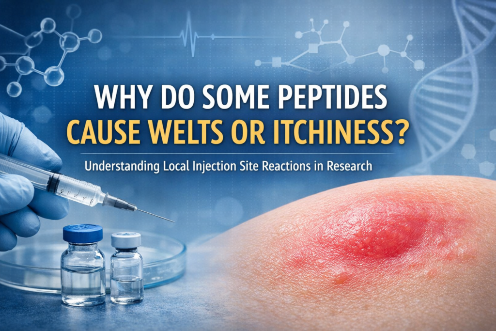
Why Do Some Peptides Cause Welts or Itchiness?
Understanding Local Injection Site Reactions in Research Settings
Localized skin reactions such as small welts, redness, or itchiness are occasionally observed in laboratory models following subcutaneous administration of peptides and related research compounds.
While these reactions may appear concerning at first, they are commonly reported in peptide research literature and are often transient. Understanding the mechanisms behind these responses can help researchers interpret observations more confidently.
This article explores why these reactions occur, what is typically considered normal, and when further evaluation may be warranted.
Why Can Peptides Trigger Welts or Itchiness?
1. Mast Cell Activation and Histamine Release
Certain peptides, particularly cationic (positively charged) or biologically active signaling peptides, have been shown in research settings to stimulate mast cell degranulation.
Mast cells release histamine and other inflammatory mediators, which may cause:
– Localized swelling
– Redness
– Itchiness
– Warmth
This response is similar to what occurs with minor skin irritants or insect bites and is generally self-limiting.
Research has demonstrated that some cationic peptides can directly trigger mast cell activation independent of classical allergic pathways (Lorenz et al., 1998). Earlier research also confirmed that human skin mast cells release histamine in response to certain stimuli (Lowman et al., 1988).
Examples Noted in Research Contexts
In laboratory and observational settings, certain compounds have been associated with a higher frequency of localized histamine-related or injection-site responses.
GHK-Cu (Copper Tripeptide-1)
As a biologically active copper-binding tripeptide involved in tissue signaling pathways, GHK-Cu may produce temporary localized redness or mild inflammatory responses in some biological models.
Melanocortin Analogues
Melanocortin receptor agonists (e.g., melanotan-class compounds) have been associated in research settings with flushing, transient welts, or mild itching. These effects are believed to be related to vasoactive and mast cell signaling pathways rather than impurity.
MOTS-c
Although not classically categorized as a histamine-stimulating peptide, MOTS-c has occasionally been noted in research environments to produce mild localized firmness or irritation. These responses are often attributed to concentration variables or tissue-level signaling effects.
NAD+ (Nicotinamide Adenine Dinucleotide)
While NAD+ is not a peptide, it is commonly handled similarly in research settings. Due to its acidic nature, localized burning or welt-like reactions have been observed in some models following subcutaneous placement. These reactions are often mechanical or pH-related rather than immune-mediated.
Not all biological models will demonstrate these effects, and variability is expected.
2. Mechanical Injection Variables
Injection-related variables can influence local response, including:
– Shallow (intradermal) placement instead of subcutaneous
– Injecting too quickly
– Larger volume in a small surface area
– Repeated use of the same anatomical site
Even sterile saline alone can produce a temporary raised bump if placed too superficially.
In many cases, what appears to be a reaction may simply be localized fluid placement within the dermal layer.
3. Concentration and Reconstitution Factors
Highly concentrated solutions may increase localized irritation potential in some biological models.
Ensuring:
– Complete dissolution
– Gentle reconstitution (avoiding vigorous shaking)
– Appropriate dilution ratios
– Proper storage conditions
may help minimize mechanical irritation during laboratory handling.
What Is Considered Normal?
In research environments, the following localized reactions are commonly observed and typically self-resolving:
– Small raised welt at the administration site
– Mild redness limited to the immediate area
– Temporary itchiness
– Slight firmness under the skin
– Resolution within several hours (occasionally up to 24 hours)
These reactions generally:
– Remain localized
– Do not progressively worsen
– Do not spread significantly
– Do not produce systemic symptoms
A transient, localized response does not automatically indicate product instability, impurity, or contamination, particularly when third-party purity verification has been performed.
When to Be Concerned
In a research context, further evaluation may be warranted if observations include:
– Rapidly expanding redness beyond the injection site
– Severe or escalating pain
– Persistent swelling lasting multiple days
– Signs of infection (increasing warmth, spreading redness, discharge)
– Systemic symptoms such as generalized hives, shortness of breath, dizziness, or facial swelling
Severe or systemic reactions should be treated as medically significant and require appropriate clinical evaluation.
Laboratory compounds should be discontinued immediately if serious reactions occur.
Does a Local Reaction Mean the Peptide Is Low Quality?
Not necessarily.
Localized histamine responses can occur even with highly purified materials. Biological variability, mast cell sensitivity, mechanical factors, pH, and concentration all influence tissue response.
At Revitalize Peptide Lab:
– All peptide materials undergo third-party HPLC testing
– Each batch meets our ≥99% purity standard prior to release
– Products are handled under strict quality control procedures
A mild, localized welt is often a biological response rather than a quality failure.
Best Practices to Minimize Local Irritation (Research Context Only)
General laboratory handling considerations may include:
– Rotating administration sites
– Avoiding repeated use of the exact same location
– Allowing alcohol to fully dry before administration
– Injecting slowly
– Ensuring complete dissolution before drawing solution
– Avoiding overly concentrated reconstitution ratios
– Avoiding overly shallow (intradermal) placement and ensuring material is delivered into the intended subcutaneous layer when applicable
Very superficial placement into the dermal layer may increase the likelihood of visible welts or raised bumps, as fluid deposited intradermally tends to remain localized and elevated rather than dispersing within subcutaneous tissue.
These observations are commonly discussed in research environments and are not medical recommendations.
Conclusion
Temporary welts or itchiness following subcutaneous administration are commonly reported in research settings.
They are often related to:
– Mast cell activation and histamine release
– Mechanical variables
– Concentration differences
– Individual biological variability
– Local pH effects (in certain compounds such as NAD+)
In most cases, these reactions are mild, localized, and self-resolving.
Understanding why they occur allows researchers to interpret observations accurately without unnecessary concern.
Important Notice
All products offered by Revitalize Peptide Lab are intended strictly for laboratory research use only.
Not for human or veterinary use.
Not for diagnostic, therapeutic, or clinical applications.
This article is provided for educational and informational purposes within a research context only and does not constitute medical advice.
Sources
Lorenz, D., Wiesner, B., Zipper, J., Winkler, A., Krause, E., Beyermann, M., Lindau, M., & Bienert, M. (1998). Cationic peptide–induced mast cell degranulation. The Journal of General Physiology, 112(5), 577–591. https://doi.org/10.1085/jgp.112.5.577
Lowman, M. A., Rees, P. H., Benyon, R. C., & Church, M. K. (1988). Histamine release from human skin mast cells. British Journal of Pharmacology, 95(4), 121–130. https://doi.org/10.1111/j.1476-5381.1988.tb11560.x